
Figure 3(A-C).
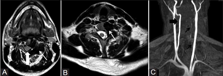
(A and B) Axial T2W MR images showing cortical hyperostosis as hypointense signal (arrow) involving left-sided hemivertebrae causing displacement of the spinal cord to the right (arrowhead) and narrowing ofleft neural foramina (arrow). (C) MR angiogram time-of-flight (TOF) image showing attenuated left vertebral artery (black arrows) and normal right vertebral artery (arrowhead)