Figure 6(A-C).

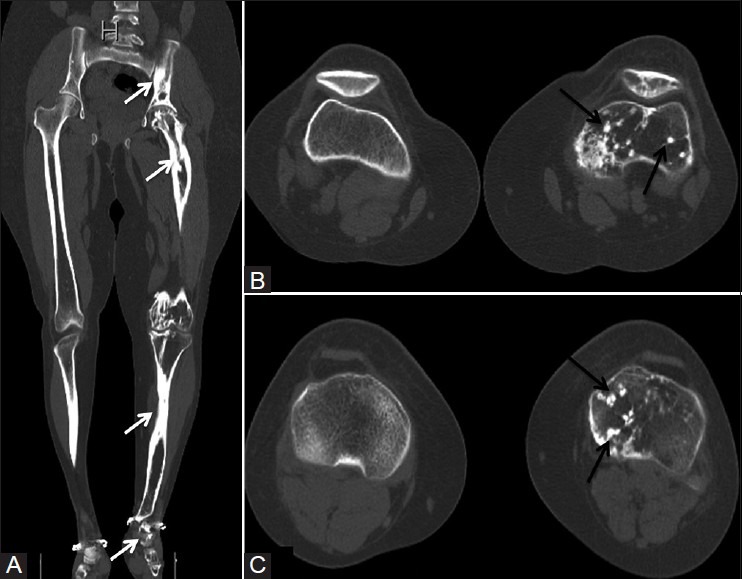
(A) Coronal reformatted CT scan image showing flowing cortical hyperostosis (white arrows) involving the left limb. (B and C) Axial CT scan images depicting multiple bone islands (black arrows) seen in femur, tibia, and patella

(A) Coronal reformatted CT scan image showing flowing cortical hyperostosis (white arrows) involving the left limb. (B and C) Axial CT scan images depicting multiple bone islands (black arrows) seen in femur, tibia, and patella