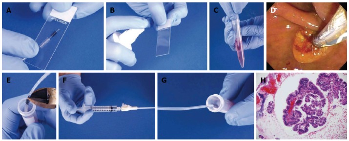
Figure 2.

Brushing technique. Two passes performed in the stricture. A, B: The first pass was used to make two smears (A), with one smear sprayed with fixative (B); C: The brush was then agitated in the RPMI cytology fluid to dislodge material into the fluid; D: The brush was rinsed with water. A second pass was performed with the same brush; E: The brush was cut off into the same tube of RPMI; F, G: Contents of catheter were flushed via salvage cytology technique; H: The sample was processed as a cell block.
