
Figure 4.
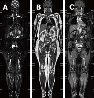
Whole-body magnetic resonance imaging of an osseous myeloma lesion. Whole-body magnetic resonance imaging: short-tau-inversion-recovery sequence (A), T1-weighted image (B) and T1-weighted image with fat suppression after contrast administration (C) showing an osseous lesion in L4 (arrows) representing an osseous myeloma manifestation.