
Figure 4.
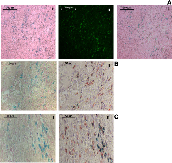
Photomicrographs of Prussian blue and CD68-immunostained sections confirming in vivo superparamagnetic iron oxide nanoparticles(SPION) labeling of macrophages.(A) Double-stained photomicrographs of (i) Prussian-blue-stained SPIONs (ii) immunofluorescent-stained macrophages using mouse anti-rat CD68 1ry antibody (as a marker of macrophages) and anti-mouse fluorescein isothiocyanate (FITC) second antibody at 20 times magnification (iii) the overlay of the two previous images. (B, C) Photomicrographs of two separate examples of (i) Prussian blue followed by (ii) CD68-immunostained sections at 40 times magnification. Sections were stained consecutively and micrographs of the same region of interest were obtained.