
Figure 1.
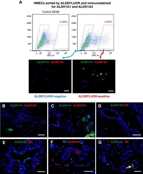
ALDH+ mammary epithelial cells do not express ER. (A) Primary human mammary epithelial cells were sorted by ALDEFLUOR (ALDE) positivity, and cytospins with ALDE+ and ALDE– cells, respectively, were stained for ALDH1A1 and ALDH1A3 isoforms. ALDE+ cells were positive for either ALDH1A1 or ALDH1A3 (lower right panel). None of these two isoforms was detectable in the ALDE– fraction (lower left panel). (B, C)In situ staining of normal breast epithelium showed that ALDH1A1 and ALDH1A3 were expressed in distinct cell populations, with no overlap. (D-G) No colocalization of ER and ALDH1A3 or ALDH1A1 was observed with immunofluorescence. Nuclear stain (blue DAPI) was performed on all sections. Scale bar = 50 μm.