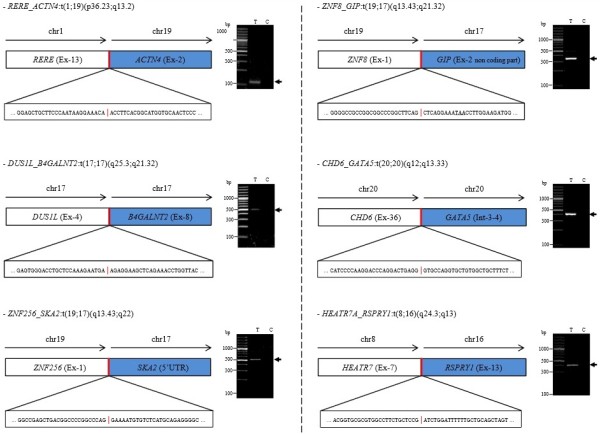
Figure 4.

RNA sequencing identification and validation of the six fusions found in invasive micropapillary carcinoma. Schematic representations of the fusions between gene 1 and gene 2 are displayed with the precise sequence of the breakpoint region. In the ZNF8_GIP fusion scheme, the underlined codon is a stop codon. RT-PCR detection of fusion genes in tumour (T) and constitutional (C) RNAs are shown on the right panel for each fusion. The numbers of split and spanning reads, together with the genomic strands of the fused genes, are given in Additional file 3: Table S8. Primer sequences used for RT-PCR validation are provided in Additional file 4: Table S9. chr, Chromosome; UTR, Untranslated region.
