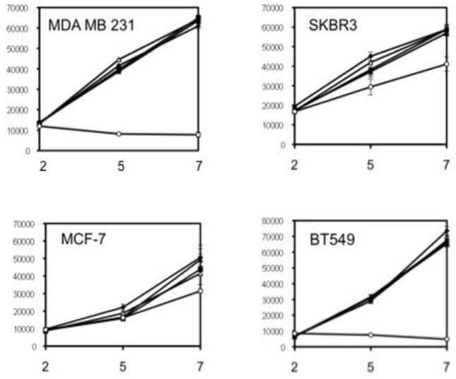
Figure 5.
Proliferation of human breast cancer cells. The cell lines MDA-MB-231, SKBR3, BT549 and MCF-7 (clockwise) were grown in culture as described with 0.5% fetal serum (open circles) or 10% fetal serum alone (closed squares) or with added EGF (50 ng/ml; closed diamonds), or SLO (2 units/ml, open triangles; 10 units/ml, closed triangles). At days 2, 5, 7 the number of viable cells was assayed by Alamar blue and fluorescence intensity measured in arbitrary units.

