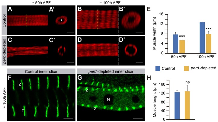
Fig. 4.
Myofibril assembly is disrupted in perd-depleted muscles. (A–D′) Confocal micrographs of muscles at 50 and 100 hours APF stained with Rhodamine–Phalloidin (red). A′–D′ show orthogonal confocal cross sections of the muscles in A–D, respectively. The asterisk marks the muscle lumen. (A–B′) From 50 to 100 hours APF, control muscles widen by increasing the amount of myofibrils. (C–D′) perd-depleted muscles are thinner than control at both 50 and 100 hours APF. (E) Quantification of the muscle width of both control and perd-depleted muscles at 50 and 100 hours APF (means±s.d., n = 27). Note the perd-depleted muscles grow very little compared to control muscles. (F,G) Longitudinal confocal cross sections of the muscles, at 100 hours APF, expressing Zasp–GFP (green) to label the Z-bands (Z). (F) In control muscles, Zasp–GFP is found in bands spanning the fiber width from the sarcolemma to the muscle lumen. (G) These bands are thicker and shorter in perd-depleted muscles. In addition, some Zasp–GFP can be seen in the cytoplasm surrounding the nuclei (N). (H) Quantification of the muscle length reveals that perd-depleted muscles are as long as controls (means±s.d., n = 27). ns, not significant; ***P<0.001. Scale bars: 5 µm.

