Fig. 5.
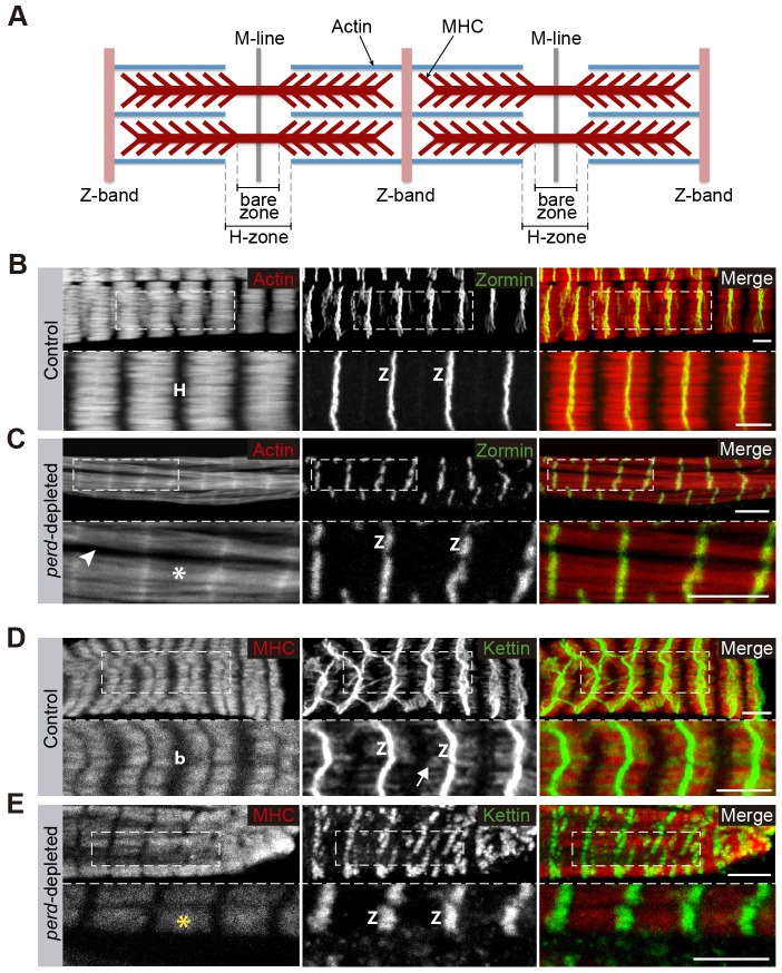
Sarcomeric structure is affected in perd-depleted muscles. (A) Schematic diagram of the sarcomeric structure. (B–E) Confocal micrographs of control (B,D) and perd-depleted muscles (C,E). (B,C) Rhodamine–Phalloidin (red) and anti-Zormin antibody (green). In control muscles, the Z-band protein Zormin forms straight lines (B); these are discontinuous and irregular in perd-depleted muscles (C). In addition, the H-zone (H), labeled by the absence of Phalloidin in controls (B), is missing in perd-depleted muscles (white asterisk in C), Phalloidin staining also shows gaps between myofibrils (arrowhead in C). (D) In control muscles, Kettin is found at the Z-bands and extends towards the central part of the sarcomere (arrow). (E) However, in perd-depleted muscles, Kettin is restricted at the Z-bands. In addition, MHC staining (red) reveals that the bare zone (b), contained within the H-zone, is also missing in perd-depleted muscles (yellow asterisk). To facilitate sarcomere visualization, the magnified regions are single slices. Z, Z-band. Scale bars: 5 µm.

