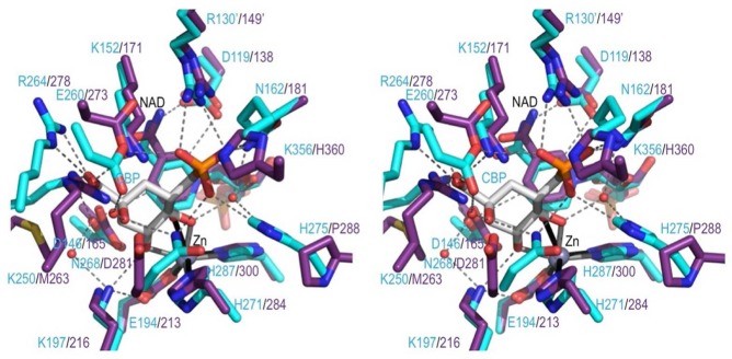
Figure 6.

Comparing the ValA active site region with the DHQS·CBP complex. Stereoview of select active site residues in ValA (purple) overlaid on the DHQS (cyan) in complex with CBP (white) shown in roughly the same orientation as DAHP in Figure 1. H-Bonding interactions in the DHQS active site (dashed lines) and coordination bonds with Zn2+ (solid lines) are shown. A prime on a residue number means it is from the other subunit of the dimer.
