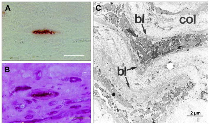
Figure 2.
Advanced human atherosclerotic plaque showing apoptotic smooth muscle cells (SMCs). A is stained by the TUNEL technique and B is the same TUNEL-labeled section subsequently stained by PAS (periodic acid-Schiff) stain (TUNEL+PAS technique). A labeled nucleus is present in a cell–poor area in the fibrous cap (A), and this TUNEL-positive nucleus belongs to a cell that is surrounded by a prominent cage of PAS-positive basal laminae (B). This points to an SMC undergoing apoptotic cell death. Most of the SMCs in this region do not express α-SMC actin. Adjacent to this cell are PAS-positive empty cages of thickened basal lamina. C is transmission electron microscopy (TEM) image of an advanced human atherosclerotic plaque. Two SMCs are demonstrated that are completely disintegrated into myriad vesicles (granulovesicular degeneration). The prominent basal laminae (bl) around these clusters of vesicles led us to conclude that the vesicles are of SMC and not of macrophage origin. (Reproduced with permission from Kockx MM, et al. Circulation. 1998;97:2307–2315.)

