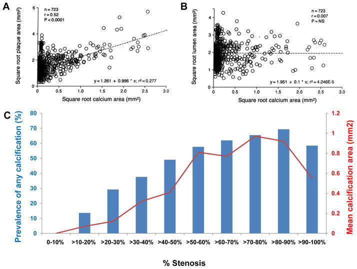
Figure 6.
A shows correlation between square root of coronary calcium area values (mm2) detected by histopathologic and microradiographic analysis and square root of plaque area values (mm2) for each of the 723 coronary artery segments in humans. B shows square root of coronary calcium area (mm2) detected by histopathologic and microradiographic analysis versus square root of lumen area (mm2) for each of the 723 coronary artery segments where no relation was identified. C shows relationship between percent stenosis and the degree of calcification in sudden coronary death victims. Each blue bar represents prevalence of any calcification (%), whereas each red dot represents mean calcification area (mm2). (A and B are reproduced with permission from Sangiorgi G, et al. J Am Coll Cardiol. 1998;31:126–133. Data in C is stratified by decades from Burke AP, et al. Herz. 2001;26:239–244)

