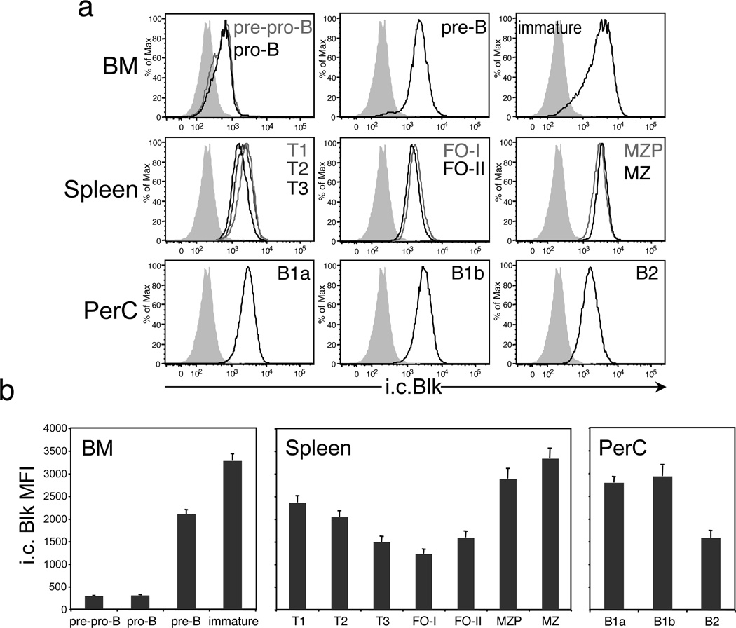
Figure 1.
Blk is differentially expressed in immature and mature B cell subsets. (a) Histograms show representative staining of the i.c. levels of Blk in gated populations of immature and mature B cell subsets in the BM, spleen and PerC of C57BL/6 mice. In the BM, these subsets include pre-pro-B (IgM− B220+ CD43+ c-Kitlo), pro-B (IgM− B220+ CD43+ c-Kit−), pre-B (IgM− B220+ CD43−), and immature (IgM+ B220lo) B cells. In the spleen, the subsets include T1 (B220+ CD93+ IgMhi CD23−), T2 (B220+ CD93+ IgMhi CD23+), T3 (B220+ CD93+ IgMlo CD23+), FO-I (CD19+ IgDhi IgMlo CD21+), FO-II (CD19+ IgDhi IgMhi CD21+), MZP (CD19+ IgDhi IgMhi CD21hi), and MZ (CD19+ IgDlo IgMhi CD21hi) B cells. In the PerC, the subsets include B1a (IgM+ CD5lo CD11b+), B1b (IgM+ CD5− CD11b+) and B2 (IgM+ CD5− CD11b−) B cells. Cells from all tissues were stained and acquired on the same day. Data are representative of two independent experiments, with 3 mice per experiment. Solid gray histograms represent Blk staining levels in mature CD4+ T cells, which express negligible levels of Blk by real-time RT-PCR analysis.7 (b) Summary of the data presented in a. Comparison of i.c. Blk levels, presented as mean fluorescence intensity values, in gated immature and mature B cell subsets.

