
Figure-5. TLR-activation increases MHC-II expression in a mouse dendritic cell line which correlates with enhanced antigen presentation.
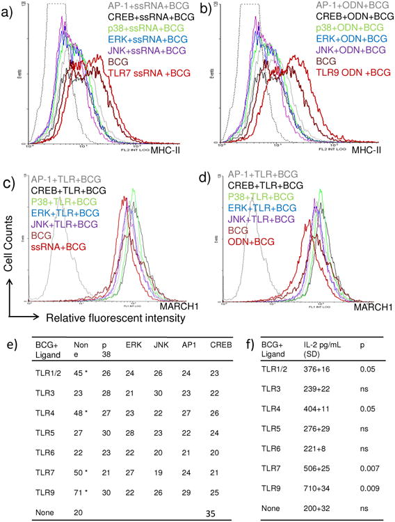
a-b) C57Bl/6 mouse bone marrow DC derived DC.2.4 cell line was activated and infected as in Fig.4. (a-b) Histograms illustrate that, TLR-7 and TLR-9 agonists enhance MHC-II and their effects are inhibited by blockade of MAPKs, AP-1 and CREB. c-d) Both agonists fail has no effect on the intracellular levels of MARCH1. e) DCs were activated with various TLR-ligands in Fig.4 and MFI values averaged from two separate experiments. Data show that ligands for TLR-9, 7, 4 and ½ enhance MHC-II. Blockade of MAPKs, AP-1 and CREB reduces expression of MHC-II or these TLR-activated DCs (* p < 0.04 vs. BCG infected DCs, t test). f) DCs were activated with various TLR-ligands in Fig.4 followed by BCG infection and IL-2 levels measured after antigen presentation. Antigen presentation by DCs is enhanced by prior activation with ligands for TLR-9, 7, 4 and 1/2 (* p values indicated for groups compared vs. BCG infected DCs, t test).