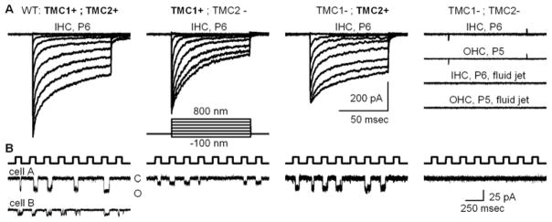
Figure 3.

Mechanotransduction currents recorded at −84 mV from hair cells of various Tmc genotypes. Genotypes are shown at the top with the protein product expressed in each case shown in bold. A, Families of whole-cell transduction currents recorded from cochlear inner hair cells at postnatal day 5 to 6 as indicated. Each family of traces is the mean of ten datasets taken from five representative cells. The stimulus was a stiff probe driven by a piezoelectrical bimorph using the step protocol shown. At the right are four representative families recorded from hair cells deficient in both Tmc1 and Tmc2. Cochlear hair cell type and age are shown for each family. The lower two families were recorded in response to 50 Hz sine wave fluid-jet deflection of inner and outer hair bundles. The scale bar applies to all current families shown in panel A. External calcium: 1.3 mM. B, Representative concatenated traces showing single-channel currents recorded from P6 inner hair cells that expressed the protein shown above panel A. The top trace shows the concatenated 100-nm square wave protocol delivered via stiff probes designed to deflect single stereocilia. For the wild-type traces (left) two examples are shown that illustrate the range of single-channel amplitudes encountered. By convention, downward deflections represent inward currents and channel openings. The scale bar at the right applies to all traces. External calcium: 50 μM. Modified from Pan et al. (2013).
