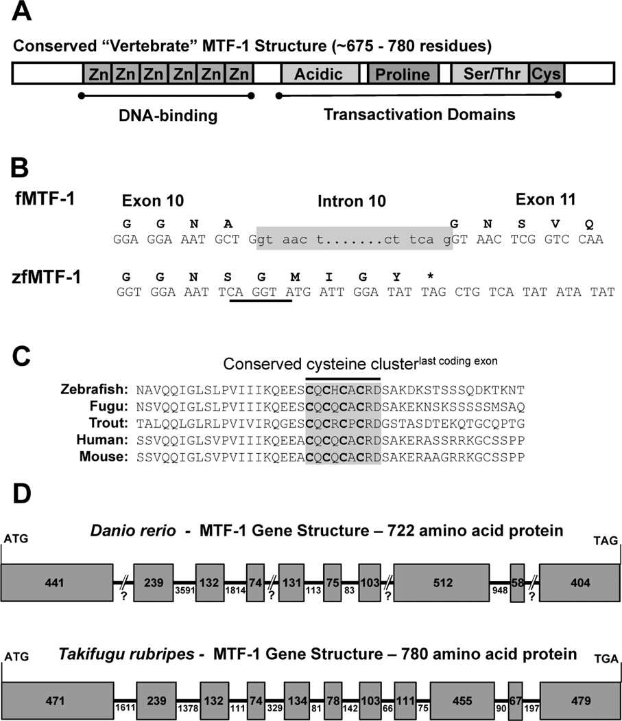
Figure 1.
Identification of a full length zebrafish MTF-1 Transcript and Corresponding Gene Structure. A) Schematic of a typical vertebrate MTF-1 protein highlighting the various conserved domains and motifs. B) Comparison of the last Takifugu MTF-1 (fMTF-1) exon/intron boundary with zfMTF-1. fMTF-1 intron 10 is highlighted in grey. Note the underlined canonical donor splice site (CAG/GTA) five codons from the putative stop codon (*) of the zfMTF-1. C) Alignment of the primary structure of the C-terminal end of various vertebrate MTF-1’s with the putative translation of the “missing” zfMTF-1 exon. The highly conserved cysteine motif is shaded in a grey box. D) A comparison of the genetic structure of the coding exons from Takifugu and zebrafish. Exons are designated by the shaded boxes and introns are represented by the lines between boxes. The numbers in the boxes or below the lines represent length in nucleotides.

