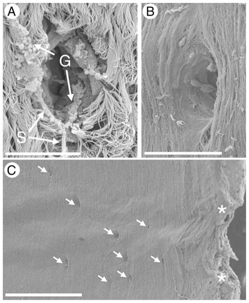
Fig. 4.
Scanning electron microscopy of gland openings. A: With surface epithelium intact. Note strands of mucus and intact mucous granules. B: With surface epithelium removed by protease digestion. C: Low power photograph showing predominant localization of gland openings between cartilaginous rings. Asterisks indicate the edges of the cartilaginous rings. Scale bars = 20 μm (A and B), 500 μm (C).

