
Figure 1.
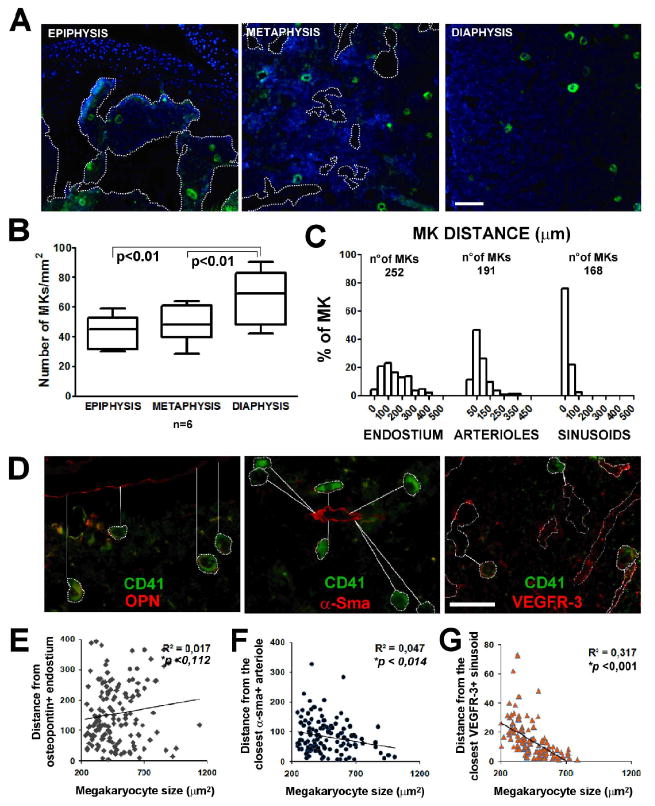
Megakaryocyte localization within the bone marrow. A) Immunofluorescence staining of CD41+ Mks (green) in bone marrow of mouse femur. Images were acquired with a 10x/0.30 Olympus UPlanF1 objective. Scale Bar=100 μm. B) Quantification of Mks in epiphysis, metaphysis and diaphysis of mouse femur. Axiovision 4.5 software was used to perform this analysis expressed as absolute number of Mks per mm2 of bone marrow surface. p value <0.01. C) Distribution of Mks related to their distance from osteopontin (OPN) positive endosteal surface, α-smooth muscle actin (α-SMA) positive arterioles and VEGFR-3 positive sinusoids within bone marrow. D) Immunofluorescence staining of CD41+ Mks (green) in proximity of OPN, α-SMA, VEGFR-3 positive structures (red). Images were acquired using 40x objective. Scale Bar=100 μm. All images were acquired using the Olympus BX51 fluorescence microscopy. E-F-G) Correlation between Mk dimension and distance from endosteal surface, arterioles and sinusoids. R, Spearman's correlation coefficient; line shows non linear regression; p, p value.