
Figure 3.
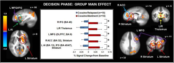
Across early and late trials, replapsed cocaine users exhibited lower activation than abstinent cocaine users in several brain regions including inferior frontal gyrus (IFG), middle frontal gyrus(MFG), anterior cingulate cortex (ACC), anterior insula (Al), thalamus, and dorsal/ventral striatum. L = left hemisphere. R = right hemisphere. BA = Brodmann Area. Error bars denote +/− standard error. Asterisks denote significant group differences. The color bar depicts normalized voxel intensity differences, with positive values indicating greater intensity in the abstinent group than the relapsed group.