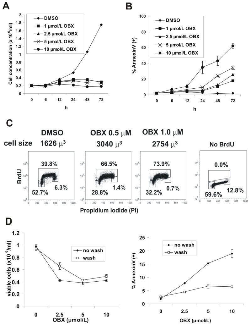
Figure 1.
Obatoclax induces apoptosis in AML. A - OCI-AML3 cells were incubated with various concentrations of obatoclax (0, 1, 2.5, 5, and 10 μM) and the number of viable cells was determined as described in the Materials and Methods. B - OCI-AML3 cells were treated with increasing concentrations of obatoclax for various times and phosphatydil serine (PS) externalization was monitored by flow cytometry by staining with Annexin V-APC. C – OCI-AML3 cells were treated with 0.5 and 1.0 μM of obatoclax for 48 h and BrdU incorporation was quantitated by flow cytometry as described in the Materials and Methods. Cell volume was determined from the average diameter measured by the ViCell XR analyzer. D – Cells were treated with obatoclax for 1 h and washed twice in serum containing media. Cells were then cultured under standard conditions for 48 h, and cell viability and apoptosis were quantitated as described in the Materials and Methods.

