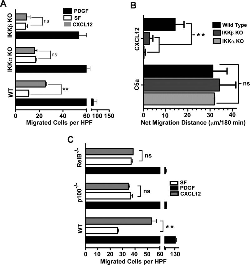
Figure 1. IKKα, IKKβ and NF-κB p52 and RelB are each required for cell migration in response to CXCL12.
(A) Chemotaxis of immortalized WT, IKKβ KO and IKKα KO MEFs (5 × 10⁴ cells) was evaluated using a 48 well microchemotaxis chambers as previously described (30). Cells were allowed to respond to CXCL12 (50 ng/ml), serum-free media (SF) as the negative control and PDGF (10 ng/ml) as the positive control for robust cell migration. The number of cells that traversed an 8 μm pore-size polycarbonate filter after a 3 hour incubation at 37°C were counted using a microscope at 400× magnification. Numbers represent mean ± SEM of cells per high power field (HPF), n = 4, ** indicates p<0.01, ns = not significant. PDGF served as a positive migration control for all 3 cell backgrounds but only to illustrate that each cell type migrate towards PDGF but in contrast to WT MEFs, IKKα KO and IKKβ KO cells do not migrate towards CXCL12. The absolute degrees of migration of WT, IKKα KO and IKKβ KO MEFs towards PDF are somewhat differ from each other, which likely reflects intrinsic properties of these different cell lines. Thus, statistical analyses of the results in Figure 1A were done for WT MEFs exposed to media vs. CXCL12: IKKα KO MEFs exposed to media vs. CXCL12; and IKKβ KO MEFs exposed to media vs. CXCL12. (B) Primary mature WT and conditional IKKα and IKKβ KO macrophages (105 cells) were exposed to CXCL12 (50 ng/ml) or C5a (2 nM) as a positive control in 48 well microchemotaxis chambers for 3 hrs as previously described (30). Data is presented as net migration distance per 400× field after subtracting basal migration in serum free media (n = 3-4). Mean value of WT macrophage basal migration towards serum free media control was 35 ± 2.6 μm. Statistical significance is indicated, ns = not significant.
(C) Chemotaxis of WT, NF-κB p100/p52−/−and NF-κB RelB−/− MEFs (5 × 10⁴ cells) was evaluated using a 48 well microchemotaxis chambers as previously described (30). Cells were allowed to respond to CXCL12 (50 ng/ml), serum-free media (SF) as the negative control and PDGF (10 ng/ml) as the positive control for robust cell migration. The number of cells that traversed an 8 μm pore-size polycarbonate filter after a 3 hour incubation at 37°C were counted using a microscope at 400× magnification. Numbers represent mean ± SEM of cells per high power field (HPF), n = 4, ** indicates p<0.01, ns = not significant.

