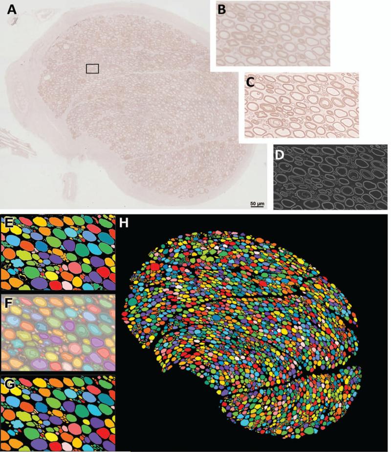
Figure 1.
Procedure for obtaining morphometric measurements in T8 motor root cross-sections. (A) Resin embedded nerves were sectioned and stained with P-phenylene diamine (PPD), imaged using light microscopy, and composite images of the entire nerve cross-sections were constructed. (B) Magnified image of area outlined in (A). (C) Images were sharpened using Photoshop™ tools. (D) Imaris® software was used to invert the image, and remove background noise. (E) Multicolored image produced by the automated count tool in MetaMorph® software. Colors indicate what software distinguishes as individual objects. Note that several adjacent axons are being mistaken for a single object. (F) Manual corrections were made in Photoshop by overlaying, and increasing the transparency of, the multicolored image over the sharpened light micrograph. (G) Multicolored images after manual corrections. (H) Corrected multicolored image of entire nerve root cross-section. (Illustration represents a T8 motor root cross section).

