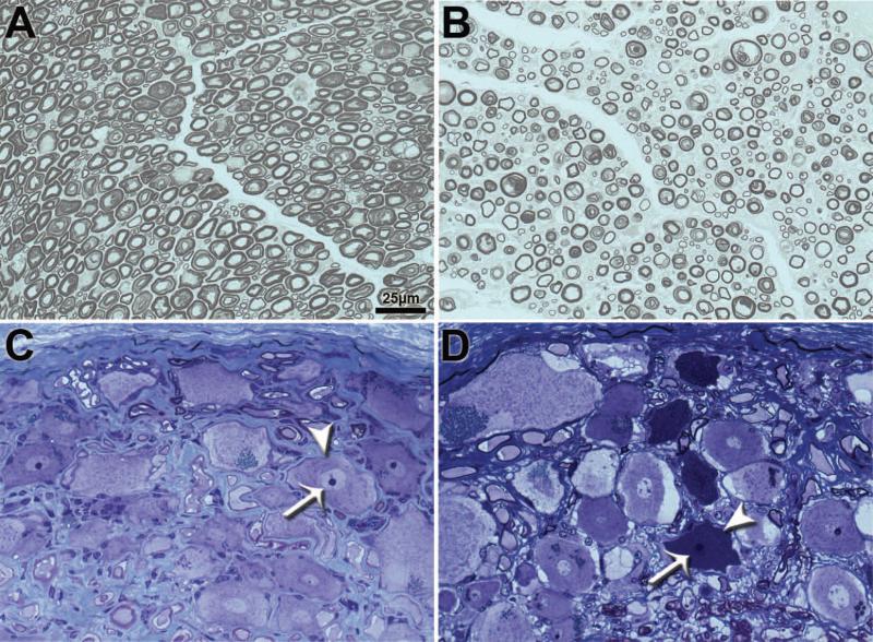
Figure 4.
Histological sections of T8 sensory roots (A and B) and dorsal root ganglia (DRG) (C and D) in PWC samples. EM-fixed sensory roots and DRG from control (A,C) and grade 4 (B,D) PWC samples were embedded in resin and stained with PPD and toluidine blue respectively. Compared to controls (A), sensory roots from grade 4 PWC (B) exhibited decreased myelinated axon density. DRG neurons from grade 4 PWC (D) contained numerous cells with condensed cytoplasm (arrowhead) and severely pyknotic nuclei (arrow) that were not present in the DRG neurons from age-matched control dogs (arrowhead and arrow in C denote cytoplasm and nucleus of normal neuron for comparison).

