
Figure 4.
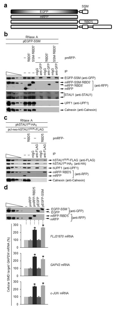
‘RBD’5 and SSM interact in trans, and expression of either inhibits hSTAU1 dimerization and SMD. (a) Diagrams of the EGFP-tagged and mRFP-tagged proteins analyzed. Unshaded regions derive from hSTAU155. (b) Western blotting using the specified antibody of HEK293T-cell lysates before (−) or after IP using anti-GFP or, to control for nonspecific IP, mouse (m)IgG (Supplementary Table 2). Lysates derived from cells (1 × 10⁷ cells per 150-mm dish) that had been transiently transfected with pEGFP-SSM (5 μg) and pmRFP (5 μg), pmRFP-‘RBD’5 (5 μg) or pmRFP-SSM-‘RBD’5 (5 μg) and subsequently treated with RNase. The four leftmost lanes analyze three-fold dilutions of lysate prior to IP. (c) As in b using lysates of HEK293T cells (1 × 10⁷) transiently transfected with pcI-neo-hSTAU155(R)-FLAG (1 μg), phSTAU155-HA3 (10 μg) and pmRFP-‘RBD5’(5 μg) or pmRFP (5 μg). (d) Western blotting (upper) and histogram of RT-PCR (lower; Supplementary Fig. 4d) of lysates of HEK293T cells (1 × 10⁷) transiently transfected with no plasmid (−) or 5 μg of pmRFP, pmRFP-‘RBD’5, pEGFP or pEGFP-SSM. After RT-PCR, the level of each cellular SMD target was normalized to the level of GAPDH mRNA, and the normalized level in the presence of no plasmid is defined as 100. All results are representative of three independently performed experiments. Error bars, s.e.m. *, n = 3, P < 0.05, determined using a one-tailed t-test.