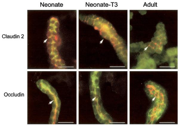
Figure 3.
Immunohistochemistry of proximal tubule expression of occludin and claudin 2. Claudin 2 and occludin were present in proximal tubules of neonate and adult rats. Neonates were treated with vehicle or thyroid hormone. (Top) Claudin 2 staining (arrow) in red. (Bottom) Occludin staining (arrow) in red. FITC-labeled LTA shows proximal tubule brush border membranes in green. Magnification: ×630. Bar = 20 μM.

