Fig. 1.
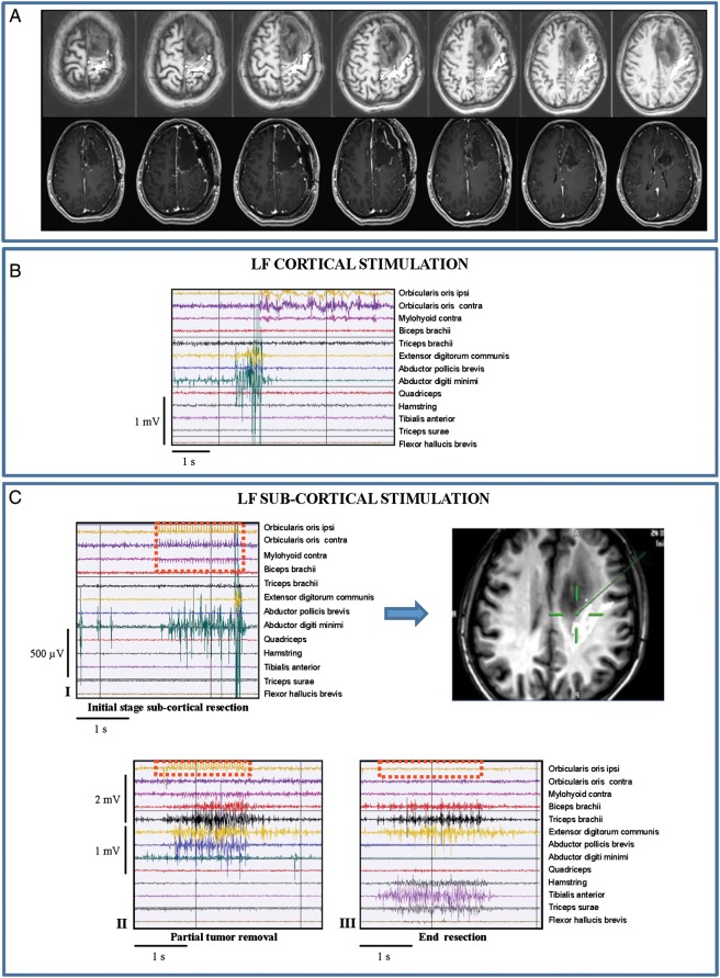
Low-frequency (LF) stimulation (60 Hz ): tumors with sharp borders (Group 1/subgroup A †and Group 2/subgroup A). Representative case: 34-year-old female with a short history of partial seizures (2 episodes of speech arrest), fully controlled by administration of one AED. MR studies showed a left (dominant) prerolandic mass resembling a low-grade glioma. She was operated under asleep-awake anesthesia for motor and language mapping. Histology documented a grade II oligodendroglioma, IDH1 mutated, MGMT methylated, codeleted. In the immediate postoperative course, she experienced transient motor (4/5) deficits, mainly at the upper limb, fully recovered. (A) Preoperative (DTI-FT reconstructions of CST, in white, were superimposed on T1-weighted images) and postoperative (postcontrast T1-weighted) MRI scan (Group 1) of the representative case. The CST was mainly displaced and slightly infiltrated. The postoperative MR showed complete resection. (B) LF stimulation of the primary motor cortex with bipolar probe-evoked EMG responses (screen shot) in distal muscles of the contralateral forearm/hand. Stimulus intensity (no stimulus artifact is visible) = 2 mA. (C) LF stimulation of the subcortical CST with bipolar probe-evoked EMG responses in different muscle groups of upper limb. Stimulation at deeper portion of CST tract (III) also involved the lower limb muscles. †Stimulus intensity = 10 mA (I and II); 6 mA (III). Stimulus artifact indicated by dashed red line.

