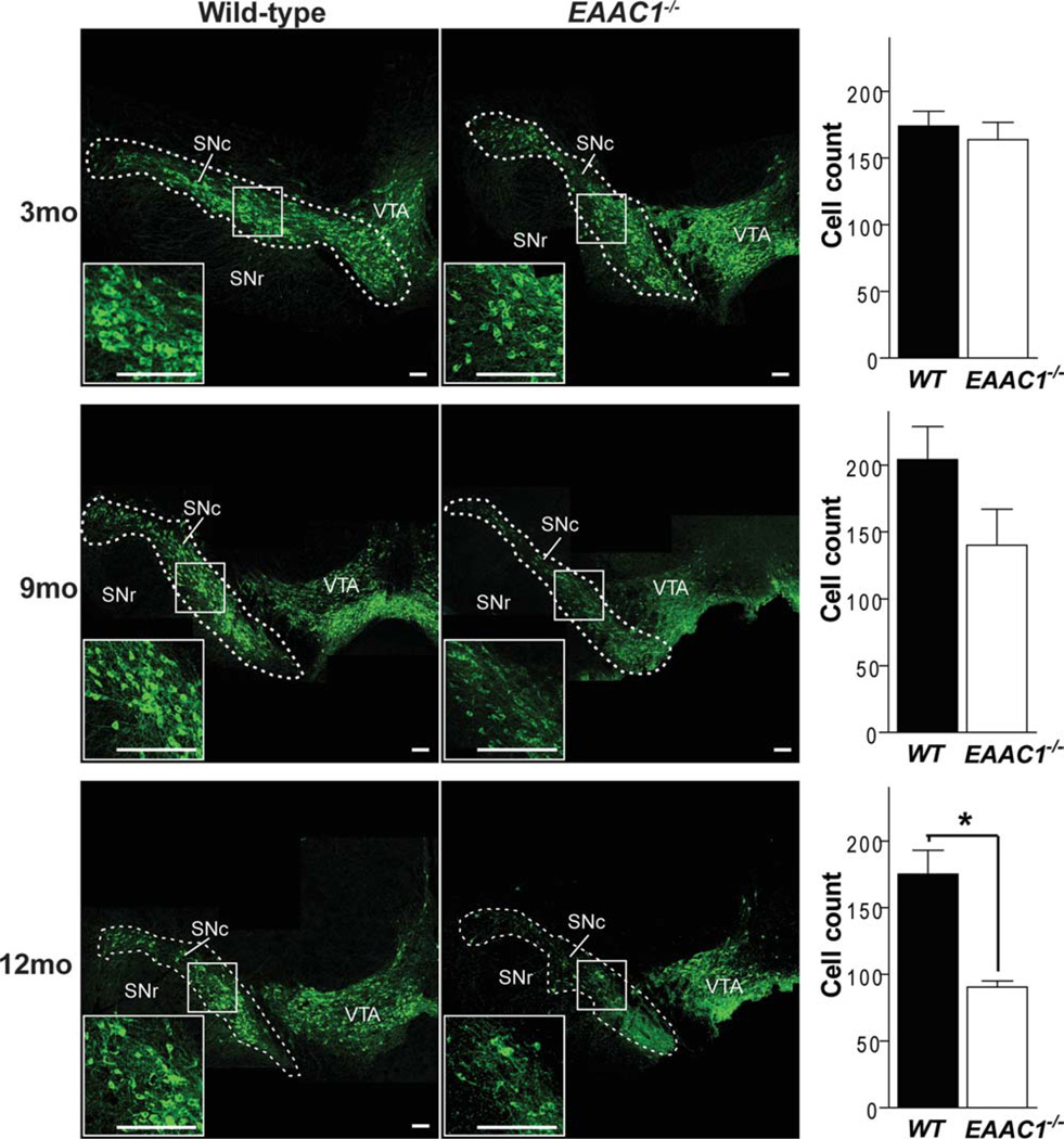
FIGURE 1.
Progressive loss of SNc dopaminergic neurons in EAAC1−/− mice. Confocal images of the dorsal midbrain of WT and EAAC1−/− mice, with dopaminergic neurons stained green (anti-TH). Insets show magnified view of boxed areas. Graphs show quantification of dopaminergic neurons in the SNc at 3, 9, and 12 months of age. Bar = 200 µm. **p < 0.01; n = 3–7. SNc = substantia nigra pars compacta; SNr = substantia nigra pars reticulata; VTA = ventral tegmental area; WT = wild-type.

