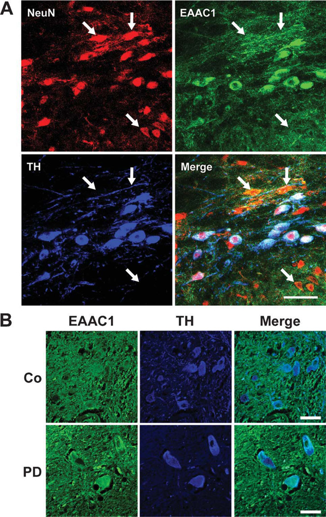
FIGURE 4.
EAAC1 expression in mouse and human SNc dopaminergic neurons. (A) Sections through mouse SNc immunostained for NeuN (red) to identify neuronal nuclei; for EAAC1 (green); and for tyrosine hydroxylase (TH, blue) to identify dopaminergic neurons. Confocal images show dopaminergic neurons to be densely stained for EAAC1, and nondopaminergic neurons (some denoted by arrows) to be less densely stained. Bar = 50µm; representative of n = 4. (B) Sections through human normal and PD brains immunostained for EAAC1 (green), and for TH (blue) to identify dopaminergic neurons. The TH-positive dopaminergic neurons show coexpression of EAAC1 in both PD and control brains. Bar = 50µm; representative of n = 4 control and 4 PD brains.

