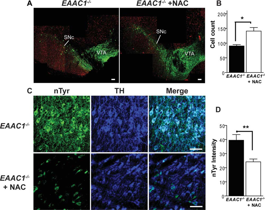
FIGURE 6.
N-acetylcysteine improves survival of SNc dopaminergic neurons in EAAC1−/− mice. (A) Images prepared as in Figure 1, with dopaminergic neurons stained green (anti-TH) and neuronal nuclei stained red (anti-NeuN). Neuronal loss in the SNc is reduced in mice treated with N-acetylcysteine (NAC). Bars = 200µm. (B) Cell counts of SNc dopaminergic neurons in EAAC1−/− mice with and without NAC treatment. **p < 0.01; n = 5–6. Note that the cell count data for EAAC1−/− mice without NAC treatment are the same as in Figure 1, reshown here to facilitate comparisons. (C) Sections are stained for nitrotyrosine (nTyr, green) as a marker of oxidative stress, and for tyrosine hydroxylase (TH, blue) to identify dopaminergic neurons. Bars = 50µm. (D) Quantified data show that the nTyr signal localized to dopaminergic neurons is reduced in the EAAC1−/− mice treated with NAC. **p < 0.01; n = 5–7.

