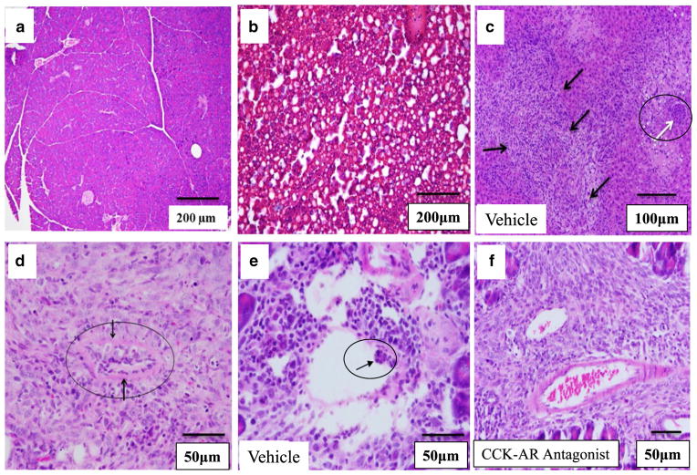
Fig. 4.
Histopathological characteristics of Panc-02 tumors and noncancerous tissues from mice. a H&E of a normal pancreas (without a tumor) from a mouse on a high-fat diet showing a lack of inflammatory or fat infiltrate. b H&E of a liver from a mouse on the high-fat diet but without a tumor (control) showing extensive steatosis. c H&E staining of a liver section from a representative tumor-bearing mouse given a high-fat diet/vehicle treatment. Multiple metastatic lesions (black arrows) and tumor emboli occluding the portal vein (white arrow) are shown. d Metastatic lesion to the mouse mesentery is shown with a tumor emboli in the center (black circle). e Tumor emboli in a small artery (arrow) from a representative vehicle-treated mouse on a high-fat diet. f Tumor vessels from a representative mouse on the high-fat diet treated with CCK-AR antagonist contain blood cells but lack tumor cell emboli. Scale bars are shown on each figure

