Figure 2.
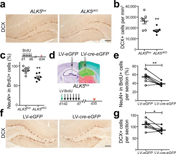
Ablation of ALK5 results in fewer newborn neurons in the dentate gyrus. (a,b) Immunohistochemical detection (a) and quantification (b) of DCX labeled cells in the dentate gyrus from 3-month-old ALK5flox and ALK5cKO mice (n = 7 mice per group, 5 sections per mouse, P = 0.0086). (c) Quantification of the percentage of BrdU and NeuN double-labeled cells in the dentate gyrus from 3-month-old ALK5flox and ALK5cKO mice after long-term BrdU labeling (n = 7 mice per group, 5 sections per mouse, P = 0.0022). Experimental design inserted on top. (d) Experimental design for the stereotaxic injection of lentivirus into the left or right dentate gyrus of ALK5flox mice at the age of 2 months and the long-term BrdU-labeling paradigm. LV, lentivirus. (e) Quantification of BrdU and NeuN double-labeled newborn neurons in the dentate gyrus from stereotaxically injected mice (n = 7 mice, 4 sections per mouse, P = 0.0059). (f,g) Immunohistochemical detection (f) and quantification (g) of DCX labeled neurons in the dentate gyrus from stereotaxically injected mice (n = 7 mice, 4 sections per mouse, P = 0.0306). Scale bar is 100 μm (a,f). Data are presented as mean ± s.e.m. Connected dots and circles in (e, g) represent left and right dentate gyrus of individual mice, bars in (e,g) represent mean values. *P < 0.05, **P < 0.01, Student's t-test (b,c); paired t-test (e,g).

