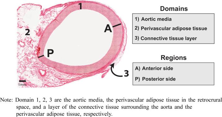
Figure 1.
Histology sample of the porcine thoracic aorta (see online version for colours)
Note: Domain 1, 2, 3 are the aortic media, the perivascular adipose tissue in the retrocrural space, and a layer of the connective tissue surrounding the aorta and the perivascular adipose tissue, respectively.

