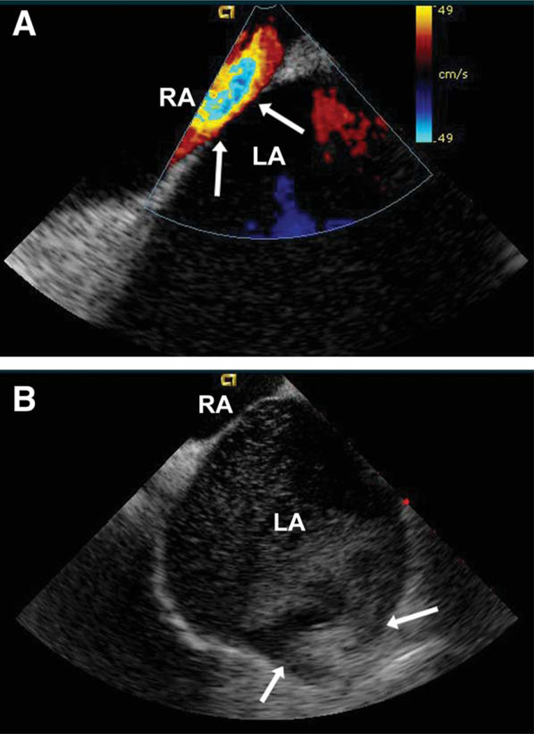
Fig. 1.
(A) Doppler ICE showing no interatrial septal defect. Arrows point to the interatrial septum. LA, left atrium; RA, right atrium. (B) Two-dimensional ICE revealing bubbles in the left atrium after injection of agitated saline in the pulmonary artery. Arrows point to the left pulmonary veins (upper and lower). LA, left atrium; RA, right atrium.

