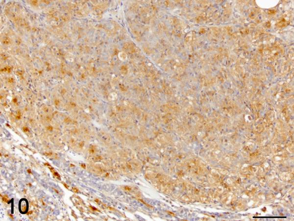
Figure 10.
Hepatocellular carcinoma. Immunolabelling TGF-β1 (brown), hematoxylin counterstain. Most neoplastic hepatocytes display moderate cytoplasmic immunoreactivity for TGF-β1. The BPDECs in the adjacent compressed hepatic parenchyma are strongly positive for TGF-β1 as in control liver. Bar = 50 μm 40X.

