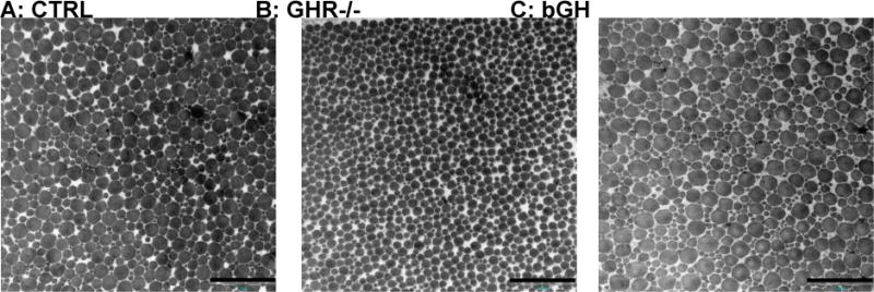
Figure 3. Representative TEM images of CTRL, GHR−/− and bGH mouse tendons.

The proximal part of the Achilles tendon was analyzed by TEM imaging in control mice (CTRL) and transgenic mice with chronic low (GHR−/−) or high GH/IGF-I activity (bGH). The three images are representative for each group. Scale bars are 500 nm.
