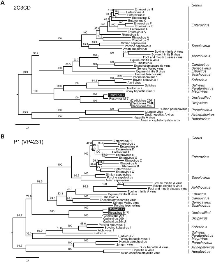
Figure 3. Rosavirus 2 is most closely-related to rosavirus M-7.
(A) Phylogenetic relationships of representative members of the Picornaviridae family were inferred from the concatenated 2C3CD amino acid alignment, generated by the maximum likelihood method. The rosavirus 2 is highlighted in black, rosavirus M-7 and cadicivirus strains are outlined. (B) Phylogenetic tree was generated from the amino acid alignment of the P1 (VP4231) region. Internal branch labels indicate the bootstrap values. The Bayesian inference method yielded trees with similar topologies.

