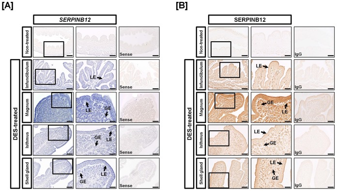
Figure 3. Localization of SERPINB12 mRNA and protein in oviducts of DES-treated and non-treated chicks.
[A] In situ hybridization analyses indicated cell-specific expression of SERPINB12 mRNA in GE and LE of the four segments of oviducts from DES-treated chicks. However, there was no expression of SERPINB12 in oviducts from control chicks. [B] Immunoreactive SERPINB12 protein was localized to LE and GE of oviducts of chicks treated with DES, especially the magnum. For the IgG control, normal rabbit IgG was substituted for the primary antibody. Sections were not counterstained. Legend: LE, luminal epithelium; GE, glandular epithelium; Scale bar represents 200 µm (the first columnar panels, sense and IgG) and 50 µm (the second columnar panels).

