
Figure 3.
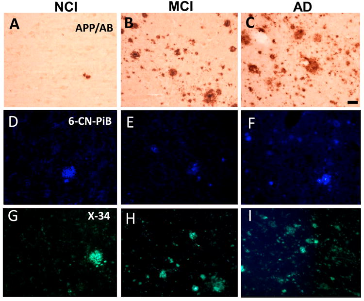
Photomicrographs of APP/Aβ (A-C), 6-CN-PiB (D-F) and X-34 (G-I) positive plaques in the precuneus in NCI, MCI and AD. Note the presence of many more APP/Aβ, 6-CN-PiB and X-34 plaques in AD (C, F and I) compared to NCI (A, D and G), while the difference with MCI (B, E and H) appeared intermediate between AD and NCI. Scale bar shown in C is same for A and B = 50 μm and in D-I = 40 μm.