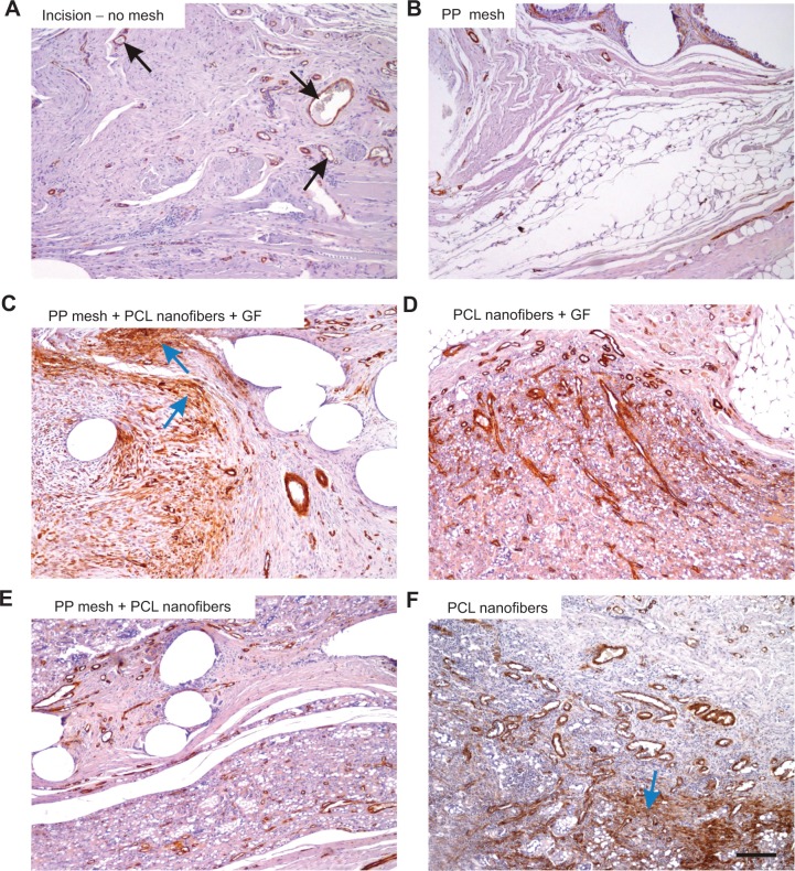
Figure 9A–F.
α-Smooth-muscle positivity in the scaffolds under study.
Notes: The density of the microvessels (some of them pointed out with black arrows) and the area fraction of actin-positive cells (vascular smooth muscle and myofibroblasts, some of the accumulated myofibroblasts highlighted with blue arrows) were highest in the PP mesh samples functionalized with PCL nanofibers enriched with adhered GF (C), followed by the PP mesh functionalized with PCL nanofibers (E), PCL nanofibers (F), and PCL nanofibers enriched with adhered GF (D), while the lowest values were found in samples of pure PP meshes (B) and sham-operated animals with no mesh (A). Immunohistochemistry for α-smooth-muscle actin, counterstaining Gill’s hematoxylin. Magnification 100×, scale bar 200 μm.
Abbreviations: PP, polypropylene; PCL, poly-ε-caprolactone; GF, growth factor.

