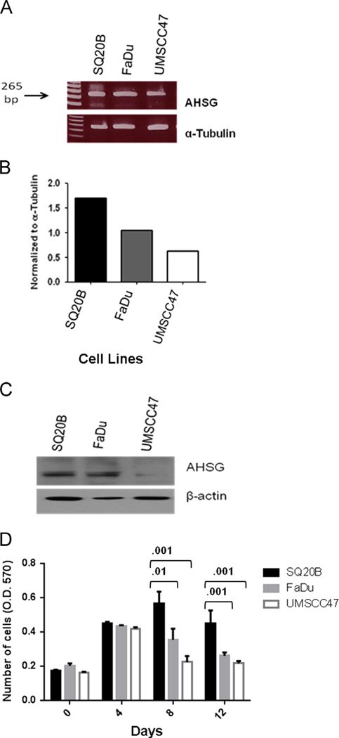
Fig. 1.
AHSG message and protein levels in HNSCC cell lines. (A) HNSCC cell lines FaDu, SQ20B and UMSCC47 were serum-starved for 48 h. Cells were washed twice with PBS. RNA was extracted using the Qiagen RNeasy Kit. Equal concentrations of each RNA sample were used to detect mRNA expression. (B) Densitometric analysis of AHSG and β-actin. AHSG relative expression was determined by normalization to β-actin. (C) The indicated HNSCC cell lines were serum-starved for 48 h. Cells were washed twice with PBS and cells were removed from the flask by scraping to ensure intact cell surface receptors were included. Protein was released from cells in RIPA buffer. Equal amounts of proteins were dissociated and analyzed in 4–12% SDS gels and immunoblotting with the indicated antibodies. (D) Cells were harvested by trypsinization, rinsed and SQ20B, FaDu and UMSCC47 were resuspended in serum-free medium. Cells (1 × 10⁴ cells/well) were seeded in SFM in 24-well plates, and incubated at 37 °C. A total of 4 plates per experiment were seeded and at days 0, 4, 8 and 12, Presto Blue was added at a ratio of 1:10 and absorbance read at 570 nm after incubation for 30 min at 37 °C to give relative viable cell numbers. Bars represent mean cell numbers ± SD from three independent experiments.

