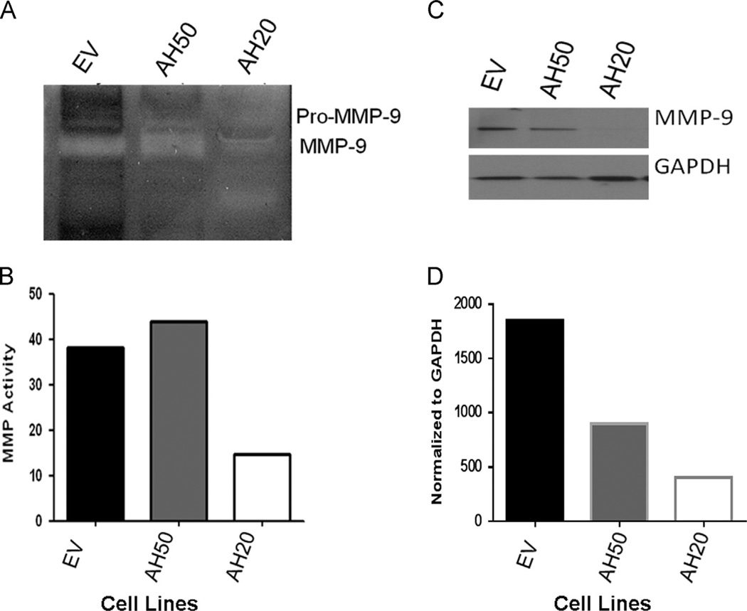
Fig. 6.
AHSG protects MMP-9 from autolytic degradation in SQ20B HNSCC sublines EV, AH50 and AH20. EV, AH50, and AH20 were cultured in serum-free media for 96 h. (A) Conditioned medium from each cell line was collected and 6 ml of each was used for ultrafiltration to a final volume of 100 µl. Equal concentrations were dissolved in zymography sample buffer and analyzed. Serum-free EV conditioned medium was used as the control. (B) Densitometric quantitation of the zymogram for MMP-9 in (A). (C) MMP- 9 protein in the concentrated conditioned medium from EV, AH50 and AH20 was quantitated by immunoblot using antibodies to MMP-9. MMP-9 bands were normalized to GAPDH and depicted in (D).

