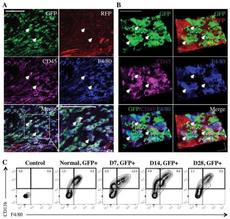
Figure 4.
Monocyte/macrophage lineage cells are the major population recruited to the wound. (A, B): Standard and three-dimensional immunohistochemical imaging of F4/80 (blue) on day 7 demonstrating that F4/80+ cells in the wound colocalize with GFP+ and CD45+ (purple) cells. White scale bar =40 μm; gray scale bar =20 μm. White arrows highlight areas of GFP+/CD45+/F4/80+ colocalization. (C): Flow cytometry of wound lysate demonstrating the GFP+/CD11b+/F4/80+ cell fraction (percentage relative to total cells) peaks at day 7 and retracts to baseline levels by day 28. Abbreviations: GFP, green fluorescent protein; RFP, red fluorescent protein.

