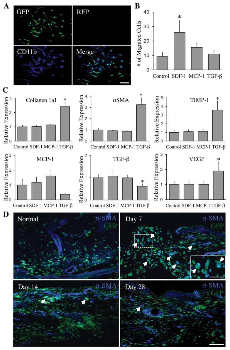
Figure 7.
SDF-1α causes bone marrow-derived CD11b+ cell migration, TGF-β1 regulates gene expression in CD11b+ cells and αSMA, the myofibroblast marker is confirmed in a subpopulation of hematopoietic cells infiltrating the wound in vivo. (A): Primary cultures of VavR bone marrow were MACS sorted for CD11b and recultured. Immunofluorescence confirmed plated cells as GFP+/CD11b+. Blue: CD11b. Scale bar =50 μm. (B): Chemotaxis assay with SDF-1α, MCP-1, and TGF-β1 shows migration of CD11b+ cells in response to SDF-1α, with *, p <.05 compared to control (n =4). (C): SDF-1α, MCP-1, and TGF-β1 treatment of CD11b+ cells shows TGF-β1 upregulating fibrosis-related genes (type I collagen, αSMA, and TIMP-1) and VEGF by qPCR. *, p <.05 compared to control (n =4). (D): αSMA is expressed in a subpopulation of infiltrating hematopoietic cells at earlier time points in the wound, but regresses with the healing of wounds. White arrows highlight areas of αSMA/GFP colocalization. White scale bar =75 μm; gray scale bar =40 μm. Abbreviations: αSMA, smooth muscle actin; GFP, green fluorescent protein; RFP, red fluorescent protein; TIMP-1, tissue inhibitor of metalloproteinase-1; VEGF, vascular endothelial growth factor.

