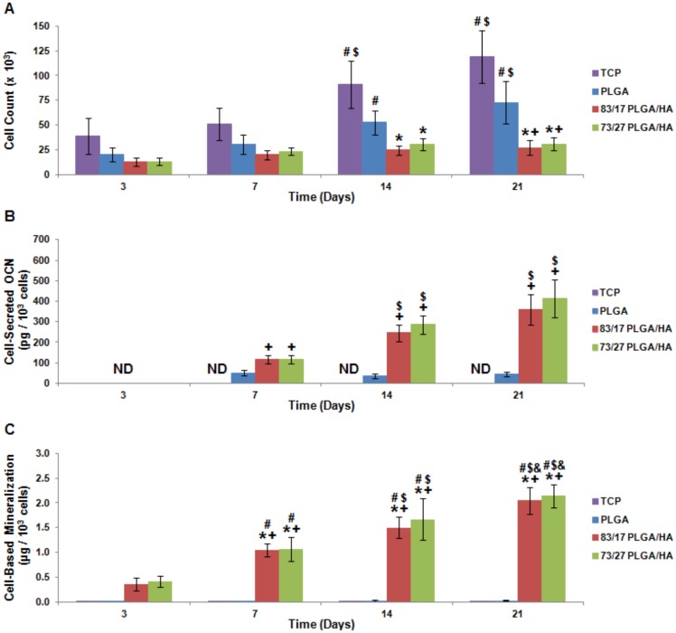
Figure 2. In vitro osteoinductivity of PLGA/HA composite scaffolds.
(A) MSC proliferation as measured by the Quant-iT PicoGreen Assay. (B) Mineralization as assessed using alizarin red staining of deposited calcium. Cell-based mineralization was quantified by subtracting scaffold-based mineralization and normalizing by cell count. (C) MSC OCN production induced by seeding on PLGA/HA scaffolds. Cell-secreted OCN as determined by ELISA and normalized by cell count. ND = Not Detectable; N = 4; p<0.05 for + = PLGA (same time point) and $ = Same Experimental Group (Day 7).

