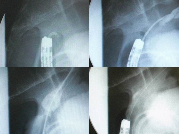
Figure 3.

Fluoroscopic findings of the procedure. Firstly, we thrust the through-the-scope thin dilator, the hardness of which was reinforced with the reversely inserted guide wire, against the center of the circular staple line to penetrate the membranous structure (upper left). When the through-the-scope thin dilator was thrust, special attention was paid not to protrude the tip of the reversely inserted guide wire from the top of the through-the-scope thin dilator because the tail of guide wire was firm and relatively sharp, and it may have stuck to and injured other organs adjacent to the anastomosis if thrust in the wrong direction. We succeeded to penetrate the through-the-scope thin dilator through the membrane (upper right). Then, the guide wire was removed from the through-the-scope thin dilator to perform fluoroscopy via the through-the-scope thin dilator. Fluoroscopy confirmed that the tip of the through-the-scope thin dilator was present in the colon oral to the anastomosis (upper right). After that, the guide wire was inserted normally through the dilator and pushed orally as possible. The fiberscope was removed and a thick dilator was inserted over the through-the-scope thin dilator through the anastomosis under fluoroscopic guidance (lower left). After removing the dilators and guide wire, a colonoscope was reinserted to apply through-the-scope hydrostatic balloon dilatation (lower right).
