Figure 5.

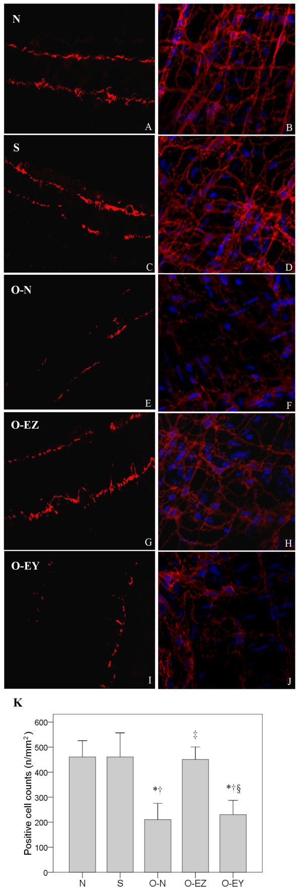
Confocal images of ICC oral to the clip labeled with DOG1 on frozen sections (A, C, E, G, I) and on whole-mount preparations (B, D, F, H, J) showing the alterations of distribution and morphology in control (A, B), sham-operated (C, D), operated without EA treatment group (E, F), operated and EA at Zusanli (G, H), operated and EA treatment at Yinglingquan (I, J). In the N and S group, DOG1-like immunoreactivity (DOG1-LI) was localized in two distinct populations of ICC (ICC-MY and ICC-DMP) within the tunica muscularis and DOG1-LI ICCs were normally present with famose processes and intact cellular networks (A-D). In the O-N group, the density of DOG1-LI ICCs were significantly reduced, the distribution was not continuous, and ICC networks were partly damaged (E, F). In the O-EZ group, an almost intact cellular networks could be observed and DOG1+ cell numbers had recovered to control values (G, H). In the O-EY group, the DOG1+ cell density was still with the same to the O-N group (I, J). Comparison between groups in positive cell counts (K). N group: Normal group; S group: Sham-operated group; O-N group: operated without EA treatment group; O-EZ group: operated and EA treatment at ST36 Zusanli point group; O-EY group: operated and EA treatment at SP9 Yinglingquan point group. *represents significant difference from N group, †represents significant difference from S group. ‡represents significant difference from O-N group. §represents significant difference from O-EZ group.
