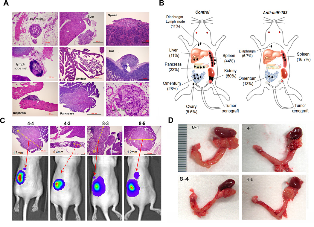
Figure 5.
The distribution and frequency of the metastatic disease in orthotopic mice of SKOV3 cells. The metastatic tumor nodules were examined and identified by histologic examination of all peritoneal organs in control and test groups of mice. A. Photomicrographs illustrated examples of the metastatic carcinomas in different anatomic sites of peritoneal organs, including the omentum, liver, spleen, pancreas, gut, diaphragm, and lymph node. B. The depicted sketch diagram illustrated the sites (indicated by labels and bar) and frequencies (in percentage) of metastatic disease in peritoneal organs in control and anti-miR-182 treated mice. C. Histology and IVIS spectrum comparison of four pancreatic metastases that were exclusively identified in control group. The actual tumor size was labeled with a yellow arrow line D. Photographs illustrated some examples of ovarian cancer extending and adhering to ipsilateral kidney that were exclusively seen in control mice.

