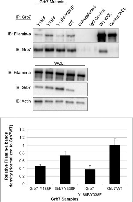
Figure 3.
The Filamin-a interaction with Grb7 tyrosine mutants is decreased. Upper panels: Western blot analysis. HeLa Cells were transfected with pCMV-FLGrb7- Y188F (full-length Grb7-Y188F mutant), pCMV-FLGrb7-Y338F, and pCMV-FLGrb7- Y188F/Y338F (double mutant) constructs. Cells were lysed and immunoprecipitated by resin-coupled rabbit polyclonal anti-Grb7 (H-70), followed by Western blot analysis with Filamin-a antibody (3 F-180) and Grb7 antibody (H-70). Whole cell lysates were also probed with the same antibodies and anti-Actin(c-2) antibody. The figure shown is a representative blot of three independent experiments, each displaying similar results. Lower panel: the results of three different immunoprecipitation experiments were analyzed by densitometry. Filamin-a band densities were divided by corresponding Grb7 mutant band densities and were shown to be normalized to Grb7 WT. Standard deviations were calculated for each sample data set and plotted in bar-graph format.

