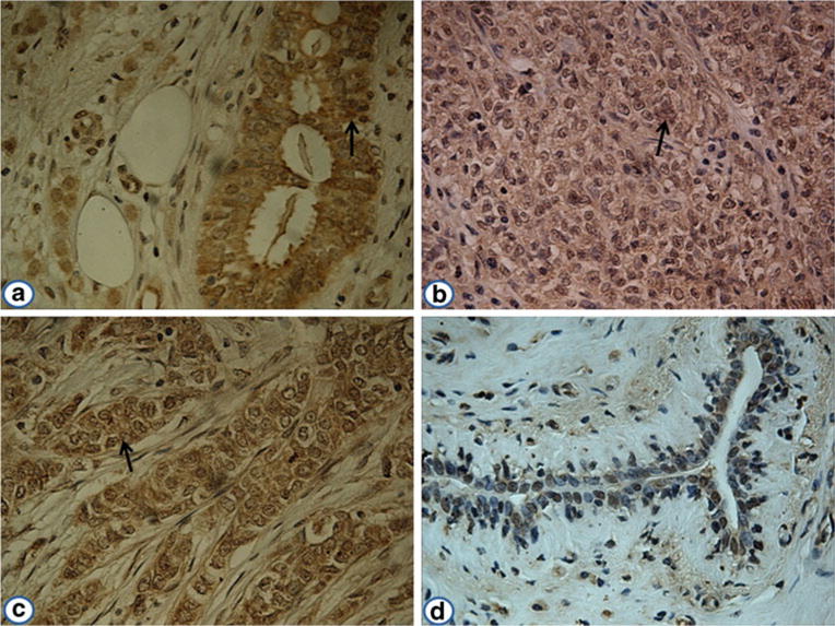
Fig. 4.

Expression of p90/CIP2A in breast cancer and adjacent normal breast tissues by immunohistochemistry. The monoclonal anti-p90/CIP2A antibody was used as primary antibody to detect the expression of p90/CIP2A in breast cancer and adjacent normal breast tissues. a Breast cancer tissue (grade I); b breast cancer tissue (grade II); c breast cancer tissue (grade III); d an adjacent normal breast tissue had negative staining (magnification, ×400). The arrows indicate p90/CIP2A positive staining
